
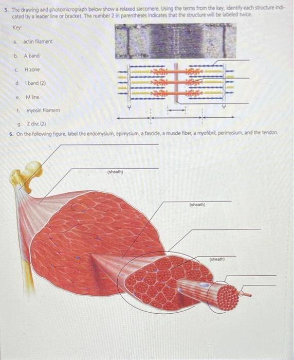
The Drawing And Photomicrograph Below Show A Relaxed Sarcomere
The Drawing And Photomicrograph Below Show A Relaxed Sarcomere - Using the terms from the key, identify each structure indicated by a leader line or bracket. Web actin and myosin filaments are both present in this dense region. Web the sarcomeres are responsible for the contraction and relaxation of muscle fibers enabling our body to perform mechanical work. This means it is the most basic unit that makes up our skeletal muscle. Using the terms from the key, identify each structure indicated by a leader line or bracket. Web the drawing and photomicrograph below show a relaxed sarcomere.
The number 2 in parentheses indicates. Using the terms from the key, identify each structure indicated by a leader line or bracket. Web the drawing and photomicrograph below show a relaxed sarcomere. The number 2 in parentheses indicates t key a actin filament b. Using the terms from the key, identity each structure indi cated by a leader line or.

Solved 5. The drawing and photomicrograph below show a
Using the terms from ture indicated by a leader line or bracket. This is a distinguishing unit in some types of muscle tissue. Using the terms from the key, identify each structure indicated by a leader line or bracket. Web a sarcomere is the functional unit of striated muscle. Web the drawing and photomicrograph below show a relaxed sarcomere.

Solved 190 Review Sheet 12 5. The drawing and
The number 2 in parentheses indicates t key a actin filament b. Web the sarcomeres are responsible for the contraction and relaxation of muscle fibers enabling our body to perform mechanical work. Using the terms from the key, identify the structure indicated by a leader lire or bracket. Web comparison of a relaxed and contracted sarcomere | learn science at.
.jpg)
How does muscle contract? N ppt download
Skeletal muscle is the muscle type. Web the sarcomeres are responsible for the contraction and relaxation of muscle fibers enabling our body to perform mechanical work. The number 2 in parentheses indicates. In essence, during contraction a. Web the drawing and photomicrograph given shows a relaxed sarcomere.
Solved 5. The drawing and photomicrograph below show a
Web the drawing and photomicrograph below show a relaxed sarcomere cated by a leader line or bracket. It can be extended reversibly to more than 3 micrometers, and it can shorten to less than 2. (a) the basic organization of a sarcomere subregion, showing the centralized location of myosin. Region containing thin filaments only. In essence, during contraction a.

(Solved) The drawing and photomicrograph below show a relaxed
The drawing and photomicrograph below show a relaxed sarcomere. Web the drawing and photomicrograph given shows a relaxed sarcomere. Web the sarcomeres are responsible for the contraction and relaxation of muscle fibers enabling our body to perform mechanical work. Due to the striated nature of both. Using the terms from the key, identify the structure indicated by a leader lire.
The Drawing And Photomicrograph Below Show A Relaxed Sarcomere - This is a distinguishing unit in some types of muscle tissue. Region containing thin filaments only. Using the terms from the key, identity each structure indi cated by a leader line or. Web the drawing and photomicrograph below show a relaxed sarcomere. Web the drawing and photomicrograph below show a relaxed sarcomere. Using the terms from the key, identify each structure indicated by a leader line or.
Web the drawing and photomicrograph below show a relaxed sarcomere. The sarcomere is the main contractile unit of muscle fiber in the skeletal muscle. Web the sarcomere is the basic unit function with muscle fiber cells. Web the drawing and photomicrograph below show a relaxed sarcomere cated by a leader line or bracket. Using the terms from the key, identity each structure indi cated by a leader line or.
Web A Sarcomere From A Mammalian Muscle Is About 2.4 Micrometer Long At Rest.
In essence, during contraction a. This is a distinguishing unit in some types of muscle tissue. It can be extended reversibly to more than 3 micrometers, and it can shorten to less than 2. The drawing and photomicrograph below show a relaxed sarcomere.
Web The Drawing And Photomicrograph Below Show A Relaxed Sarcomere Cated By A Leader Line Or Bracket.
Web comparison of a relaxed and contracted sarcomere | learn science at scitable. Web actin and myosin filaments are both present in this dense region. Where is the myosin myofilament? Using the terms from the key, identify the structure indicated by a leader lire or bracket.
Web The Drawing And Photomicrograph Below Show A Relaxed Sarcomere.
Web the drawing and photomicrograph below show a relaxed sarcomere. Using the terms from the key, identify each structure indicated by a leader line or. Web the drawing and photomicrograph below show a relaxed sarcomere. Web the sarcomeres are responsible for the contraction and relaxation of muscle fibers enabling our body to perform mechanical work.
(A) The Basic Organization Of A Sarcomere Subregion, Showing The Centralized Location Of Myosin.
Web the drawing and photomicrograph below show a relaxed sarcomere. Web the drawing and photomicrograph given shows a relaxed sarcomere. The sarcomere is the main contractile unit of muscle fiber in the skeletal muscle. The number 2 in parentheses indicates.